Intraoral scanners
Castellini intraoral scanners offer advanced technology for accurate and fast scanning of the dental arches. Innovative solutions designed to improve diagnostic efficiency and patient comfort, ensuring accurate results and optimal management of dental treatments.
Intraoral scanner
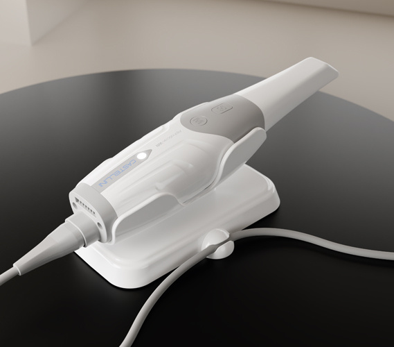
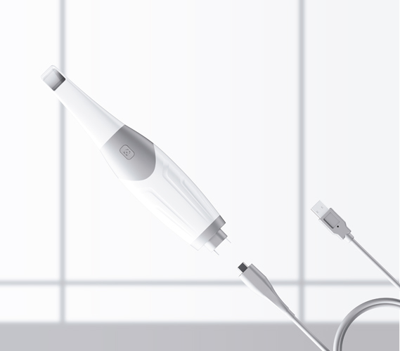
AlphaScan WR
AlphaScan WR is the wired 3D intraoral scanner designed to offer fast, accurate and user-friendly scans. With a resolution of 20 µm and a depth of field of 18 mm, it provides high quality images for detailed diagnosis, while the ergonomic, lightweight handpiece (175 g) ensures excellent handling.
Advanced integration
Integration with AlphaScan Connect optimises workflow through automatic cloud synchronisation and instant data sharing with the lab. The ScanPro software, enhanced by artificial intelligence, automatically eliminates artefacts and soft tissue, improving acquisition accuracy.
Clinical versatility
Suitable for numerous clinical applications, from implantology to orthodontics, AlphaScan WR integrates advanced tools such as Smile Design and Ortho Simulation. It also offers an APP Store to download and update applications dedicated to digital modelling and image analysis.
Optimised workflow
With a smooth workflow, cloud management and realistic images for optimal patient communication, AlphaScan WR is the ideal solution for a modern, digital dental practice.
Ergonomics and comfort
Always ready for use as it does not require calibration, it reduces set-up time and ensures smooth, seamless scans. The light and ergonomic handpiece (175 g) minimises fatigue, while the internal gyroscope allows you to navigate through the images without taking your attention away from the patient.
Precise image acquisition
The dual scanning button ensures accurate and instant image acquisition, enhancing smooth operation. With AlphaScan WR, dental practices offer an advanced clinical experience, combining state-of-the-art technology, diagnostic speed and maximum patient comfort.
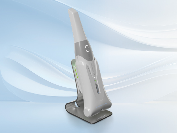
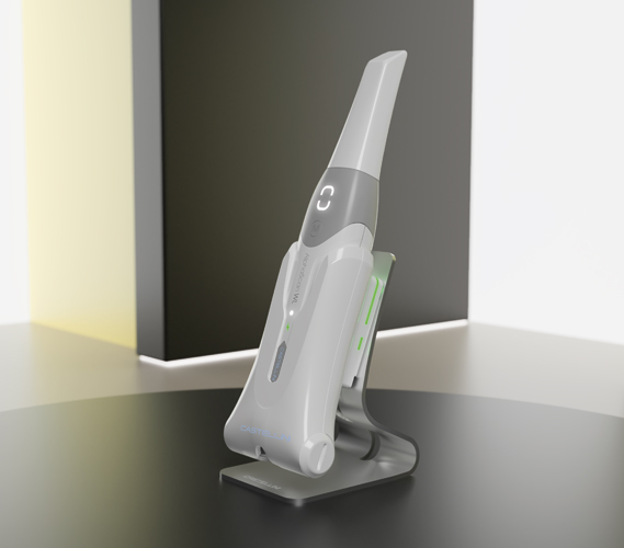
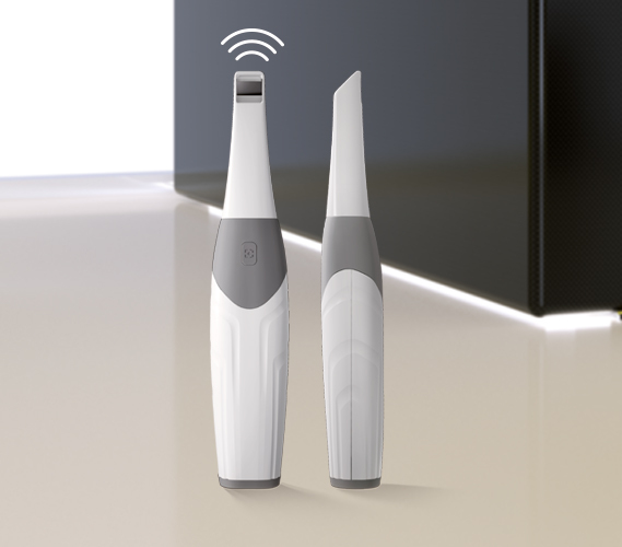
Intraoral scanner
AlphaScan WL
AlphaScan WL is Castellini wireless intraoral scanner designed to offer fast and accurate scans without limitations. With a resolution of 20 µm and a depth of field of 18 mm, it ensures high quality images for detailed diagnosis, while the ergonomic handpiece (175 g) guarantees maximum freedom of movement and both operator and patient comfort.
Cloud integration
Integration with AlphaScan Connect optimises workflow through automatic cloud synchronisation and instant data sharing with the lab. The ScanPro software, enhanced by artificial intelligence, automatically eliminates artefacts and soft tissue, improving image acquisition quality.
Advanced instruments
Perfect for various clinical applications, from implantology to orthodontics, AlphaScan WL offers advanced tools such as Smile Design and Ortho Simulation, and an integrated APP Store to update and download new features.
Digital workflow
With a smooth digital workflow, wireless connection and fast charging with a backup battery, AlphaScan WL brings the dental practice into the digital age, improving operational efficiency and the quality of diagnoses.
Optimal comfort
As calibration is not required, AlphaScan WL is always ready to use, thus reducing preparation time. The ergonomic design and balanced weight ensure a stable grip and comfortable use, reducing operator fatigue.
Smooth workflow
The integrated gyroscope allows you to control the scanner like a digital mouse, navigating between images without having to interact with the computer. Furthermore, the double scan button allows you to manage image acquisition using the same finger, regardless of the orientation of the handpiece, thus enhancing smooth operation.
Wireless technology
The wireless power supply and the back-up battery ensure continuous operation throughout the day. The handpiece automatically goes into sleep mode when not in use, and is instantly reactivated at the first movement, ensuring a seamless experience.
.png?width=2000&height=666&name=Banner%20download%20%20Castellini%20(2).png "Banner download Castellini (2)")