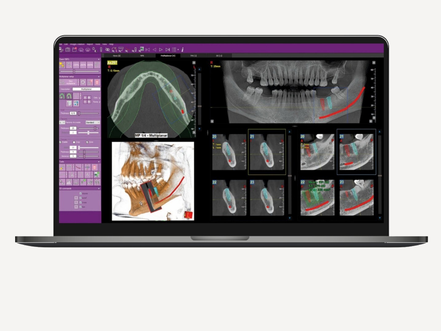
Software
Castellini iRYS

iRYS

The all-in-one software for 2D and 3D imaging that redefines efficiency. iRYS is certified for data protection and IHE-compliant with DICOM networks.

Characteristics

Characteristics

iRYS offers a comprehensive suite of features for viewing, processing and sharing acquired scans. Access all these functions directly from the dedicated workstation or from the practice’s computers.

Dose Book Tool

Improve efficiency in the collection and management of information on acquired scans: this feature enables the automatic extraction and storage of patient dose data and other information for each scan.

You can easily select scan-related information and filter the patient list by category or date. Finally, you can export the data in .pdf or .csv format for faster sharing.

Castellini has chosen to integrate Dentaldose to provide you with the best solution for managing your diagnostic exposure data.

Multi-Desktop 3D/2D

The iRYS Multi-Desktop system allows you to switch between different views, from 2D to 3D, with realistic rendering and multi-planar analysis. Everything you need for top-quality diagnostics.

Preloaded implant libraries

iRYS facilitates the selection and positioning of implants chosen among those contained in its extended library. It is also possible to change them or add new ones in just a few simple steps.

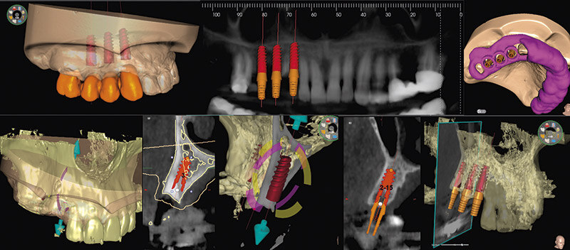
Diagnosis and planning

iRYS simplifies and optimises diagnosis and planning thanks to state-of-the-art protocols and filters.
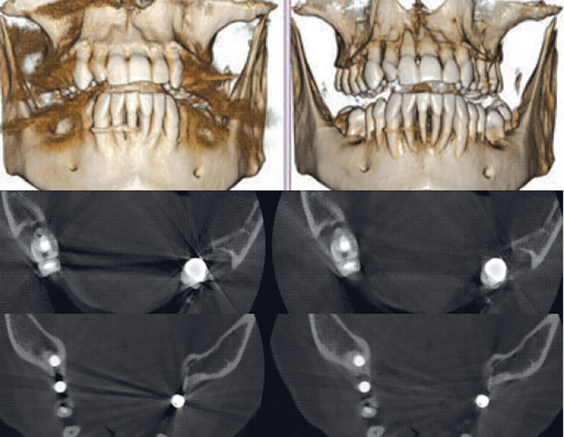
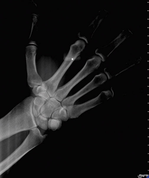
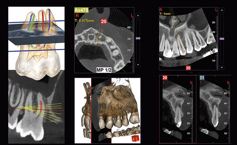
iRYS_diagnosis-and-planning_3d-smart

3D SMART

The intelligent 3D SMART (Streak Metal Artifacts Reduction Technology) feature reduces the presence of metal-caused artifacts in 3D volumes through a completely automatic procedure. Make your volumetric images usable at all times, also in the presence of implants and amalgam restorations.
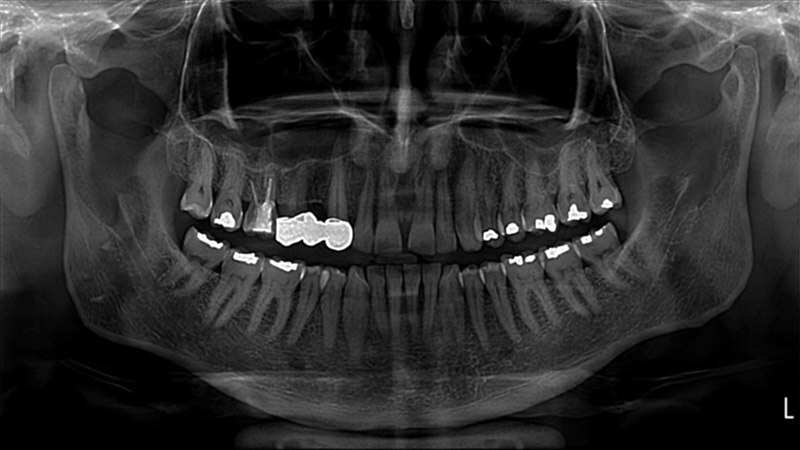
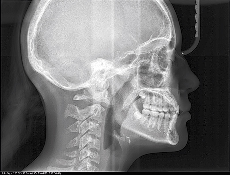
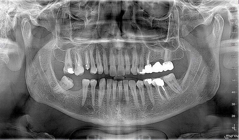
iRYS_diagnosis-and-planning_2d-pie

2D PiE

The advanced 2D PiE (Powerful image Enhancer) filters allow to maximise 2D image rendering by automatically and selectively optimising the display of different anatomical regions and by making every acquisition detail clearer, from multiple panoramic images to dentition.
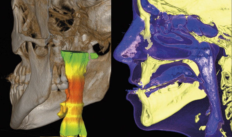
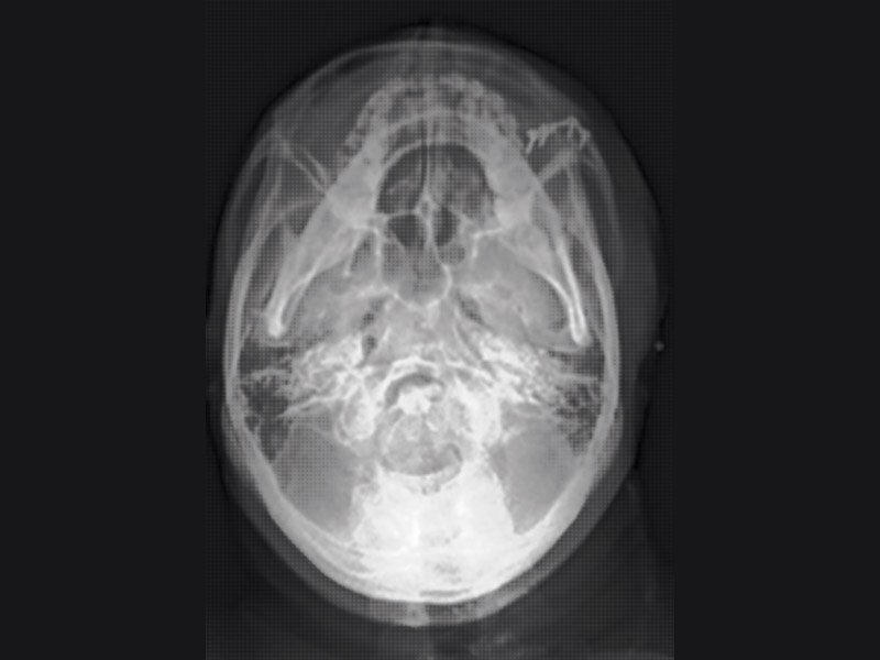
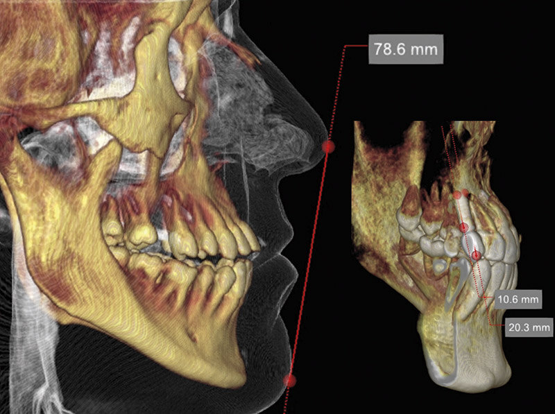
iRYS_diagnosis-and-planning_volume_of-airways

Airway volume

iRYS allows you to evaluate the upper airways volume in order to investigate possible disorders in the ENT district. This feature is also particularly useful to plan sinus lift surgery in the event of zygomatic implants or for the preliminary assessment of obstructive sleep apnea (OSA).
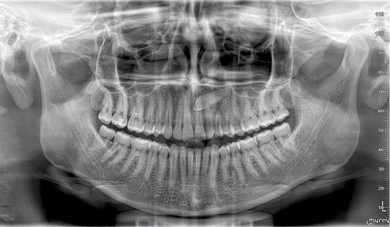
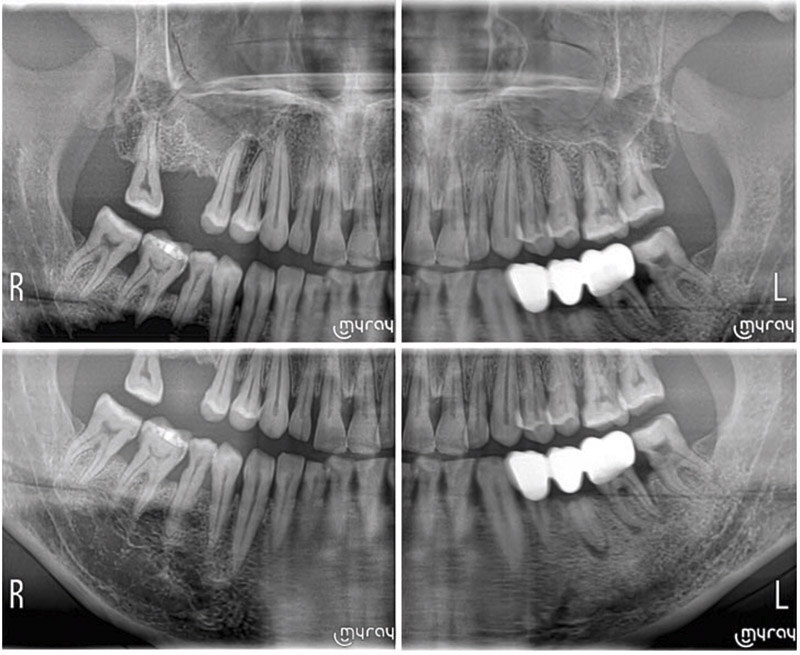
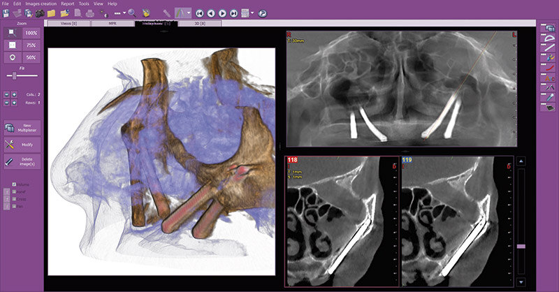
Clinical cases

iRYS_cases-clinical-2d_teleradiography_body

Teleradiography: Carpus

Accurate assessment of residual growth, with dedicated support to ensure reliable results.
iRYS_clinical cases-2d_latero-lateral_teleradiography

Teleradiography: Latero-Lateral

Detailed visualisation of bones and soft tissues, essential for high-precision cephalometric studies.
iRYS_cases-clinical-2d_teleradiography_subment-vertex

Teleradiography: Sub-mento vertex

A detailed examination of the nasal cavities, ethmoid cells and sphenoid sinuses to ensure a comprehensive diagnosis.
iRYS_cases-clinical-2d_orthogonal_panoramic

Orthogonal panoramic X-rays

Minimisation of dental overlap for optimal and detailed periodontal analysis.
iRYS_cases-clinical-2d_orthopanoramic_fast

Fast panoramic X-rays

Low radiation dose and rapid scanning, ideal for initial assessments, follow-ups and uncooperative patients.
iRYS_clinical cases-2d_orthopanoramic_definition-complete-a-quadrants

Complete detention divided into quadrants

Targeted examinations with selectable segmentation, for a reduced and controlled radiation dose.
iRYS_cases-clinical-3D_analysis-volumetric

Volumetric analyses

Obtain a precise calculation of the maxillary sinus lift volume for safe and accurate surgical planning, and direct tracing on the patient’s virtual model for detailed 3D morphological assessment.
iRYS_clinical-cases-3D_evaluation-zigomatic-plants

Assessment of zygomatic implants

Volumes with 13 x 8 cm or 13 x 10 cm FOV are the perfect tool for zygomatic implant planning as the 13cm diameter is the only one that makes it possible to include the entire zygomatic arch, without cuts.
iRYS_cases-clinical-3D_investigation-endodontics

Endodontic examination

Achieve superior diagnostics with exceptional resolution of 68 μm (exclusive to the X-RADiUS TRiO PLUS) and detailed visualisation of the mandibular canal, microfractures and root resorption.
iRYS_cases-clinical-3D_planning-implant-advanced

Advanced implant planning

Position the equipment directly on the 3D model, combine it with the STL data from intraoral scanners and define the final prosthetic project.

This allows you to plan your treatment with confidence, thanks to information on bone volume and the distance from anatomical structures (such as the mandibular canal); all whilst defining the minimum safety distance.